Mimp localization in mitochondrial cells

Research

Jun 22nd, 2015
Met Proto-Oncogene and its Ligand, HGF/SF and Breast Cancer

Prof. Ilan Tsarfaty, Ph.D.
Department of Clinical Microbiology and Immunology

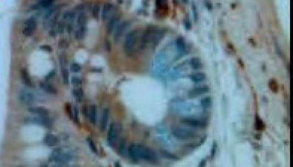
Breast cancer is the most common malignant disease in western women. In the majority of cases the cause of death in cancer patients is not the primary tumors, but complications derived from metastases at distant sites. The met proto-oncogene product (Met - a receptor tyrosine kinase) and its ligand, hepatocyte growth factor/scatter factor (HGF/SF), mediate cell motility and proliferation in vitro and tumorigenicity, angiogenesis and metastasis in vivo. Mimp/Mtch2, a mitochondrial carrier homologue cloned in our lab, is induced by Met-HGF/SF signaling and is involved in metabolic and bioenergetic processes. We have previously shown that activation of Met by HGF/SF induces an increase in tumor blood volume in a dose-dependent manner. Mimp/Mtch2 reduces cells proliferation in vitro and tumor growth in vivo. Several anti-Met targeted therapies are in development and some have entered phase III clinical trials.


The goal of our studies is to further understand the role of Met-Mimp/Mtch2 in cancer progression and metastasis, and to develop modalities for personalizing targeted Met therapy. Fluorescent tagged–Met proteins were used to study Met mitogenic effect on cells. Met induced cell motility is mediated by the formation of membrane structures such as ruffles, pseudopodia and blebs. Over expression of GFP-Met WT results in its constitutive activation, cell rounding and detachment, and dynamic nonapoptotic membrane blebbing. Bleb retraction results in numerous membrane microspikes where CFP-Met WT, YFP-actin and membrane markers accumulate.


Expression of Dominant-Negative (DN) YFP-Met alone did not induce any membrane blebbing, and co-expression of CFP-Met WT and YFP-Met DN significantly reduces membrane blebbing. Using confocal based molecular imaging we also show that Mimp/Mtch2 reduces the levels of reactive oxygen species ROS and prevents the HGF/SF induced increase in ROS. Mimp/Mtch2 also reduces the polarization of the mitochondrial membrane potential.


Mouse cerebellum

Research

Jun 22nd, 2015
The ATM-Mediated DNA Damage Response

Prof. Yosef Shiloh, Ph.D.
Department of Human Molecular Genetics and Biochemistry

Our laboratory investigates the cellular DNA damage response. This research stems from our interest in the human genetic disorder ataxia-telangiectasia (A-T), in which a central axis of the DNA damage response is missing.


Genetic defects in the DNA damage response lead to genomic instability syndromes, which usually include tissue degeneration, cancer predisposition, and sensitivity to specific DNA damaging agents. A prototype genomic instability syndrome is A-T. The disease is characterized by neuronal degeneration, immunodeficiency, chromosomal instability, sensitivity to ionizing radiation, and cancer predisposition.


Our lab has been investigating A-T since its establishment in 1985. In 1995, after 8 years of intensive work, we identified the gene that is defective (mutated) in A-T patients and called it ATM (A-T, Mutated). We went on to study the activity of its product, the ATM protein, which turned out to be an enzyme with an activity alled “protein kinase." Our current research is aimed at a broader understanding of the ATM-mediated DNA damage response. Particular attention is paid to the molecular and physiological basis of A-T, which may eventually lead to new treatment modalities for the disease. We investigate this system with cell biology methods, gene targeting in mice, and systems biology strategies including high-throughput screens, advanced proteomics and bioinformatics. A study is underway aimed at understanding the DNA damage response in the part of the brain called the cerebellum, which is badly damaged in A-T patients. Another project is searching for a drug treatment for A-T patients based on our recent understanding of the disease.

angiogenic switch

Research

Jun 22nd, 2015
Angiogenic Switch Using Rationally-Designed Theranostic Nanomedicines

Prof. Ronit Satchi-Fainaro, Ph.D.
Department of Physiology and Pharmacology

Our research interests include investigations relating to tumor biology, tumor dormancy, mechanism of action of angiogenesis inhibitors, self-assembly of polymeric architectures and novel approaches to target cancer. Throughout, we have maintained an interest in understanding the biological rationale for the design of polymer therapeutics suitable for transfer into clinical testing.


Our primary interests are the molecular basis of tumor angiogenesis and the rational design of polymer therapeutics. Our research includes identification and characterization of genes and microRNAs associated with the switch from a dormant avascular tumor phenotype to a fast-growing angiogenic tumor in human cancers and their corresponding mouse models. We focus on the design and characterization of novel drug delivery platforms, including dendrimers and hyperbranched polymer–based nanoparticles, and the design of highly-selective targeting molecules integrating biology, chemistry, protein engineering, computational approaches, material sciences
and nanotechnology to selectively guide drugs into pathological sites.


Our vision is that novel approaches to target anticancer, anti-angiogenic drugs, miRNA and siRNAs to endothelial and tumor cells to potentially treat angiogenesis-dependent diseases could transform cancer into a chronicallymanageable disease. Research methods used include sequencing, gene cloning, quantitative RTPCR, immunofluorescence, cell culture, scanning electron microscopy, mass spectrometry, NMR, HPLC, in situ hybridization, bioinformatics, polymer chemistry, molecular imaging, angiogenesis assays, animal models of cancer (human xenografts in mice, syngeneic and transgenic mice models),
pharmacokinetics and pharmacodynamics.


Carboxypeptidase E (CPE), a novel Wnt inhibitor

Research

Jun 22nd, 2015
The Wnt Signaling Pathway and Colorectal Cancer

Prof. Rosin-Arbesfeld, Ph.D.
Department of Clinical Microbiology and Immunology

The Wnt signaling pathway is involved in virtually every aspect of human development, as well as in adult homeostasis. Hyperactivation of this pathway has been linked to a wide range of cancers and especially colorectal cancer. Our aim is to understand the molecular events underlying Wnt signal transduction, as well as develop novel therapeutic strategies to fight colorectal cancer.


Current projects in the lab include:

  1. Identifying and characterizing new Wnt signaling components.
  2. Developing new anti-colorectal cancer treatment strategies.
3D reconstruction of a single red blood cell

Research

Jun 22nd, 2015
Interaction of Nanomaterials and Electromagnetic Fields with Cells

Prof. Rafi Korenstein, Ph.D.
Department of Physiology and Pharmacology

The research activity addresses the following lines of research:

  • Absorption and uptake of nanoparticles by cells in relation to drug delivery and toxicity
  • Enhancement of uptake by electrical and chemical means 
  • Treatment of cancer by electrochemical based approach
  • Assessment of genetic and epigenetic risks following in-vitro exposure to electromagnetic fields associated with cell phone communication.
  • Physiological regulation and underlying mechanism of cell membrane-cortical skeleton nanoscale mechanical fluctuations.

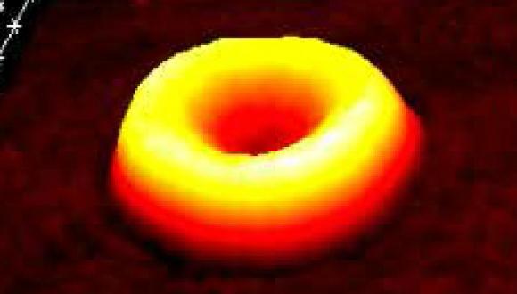
Research methods used include routine cell biology and biochemical methodologies with emphasis on special cutting edge light microscopies possessing nanometric resolution such as Digital Holographic Microscopy.


Prof. Yona Keisari

Research

Jun 22nd, 2015
Development of Cancer Treatments Integrating Radiotherapy or Electrochemical

Prof. Yona Keisari, Ph.D.
Department of Clinical Microbiology and Immunology

Cancer is currently the most devastating chronic disease affecting humankind. Today solid malignant tumors are mainly treated by surgery and/or radiotherapy to eradicate the local primary lesion, and chemotherapy, that is administered mainly to destroy remaining local or distant malignant cells. In spite of the advancement in preventing and treating cancer, morbidity and mortality remain high, especially in cases when tumors are highly metastatic, or cannot be completely removed. The main goal of our research projects is to develop in situ tumor ablation treatments of primary tumors and incorporate them with systemic chemotherapy and immuno-stimulatory agents, into combined treatment protocols.


In order to achieve efficient primary tumor ablation we developed two novel and powerful treatment modalities for solid cancer, which can be used instead or in combination with surgery. The first treatment, developed with Prof. Rafi Korenstein (Dept. Physiology & Pharmacology), is base on the use of intratumoral unipolar pulsed electric currents for the ablation (ECTA) of solid primary tumors. ECTA can be enforced by the concomitant use of chemotherapeutic agents in the treatment of tumors. The second cancer treatment, developed with Prof. Itzhak Kelson (School of Physics & Astronomy), is based on insertion into the tumor of radioactive wires that spread in the tumor alpha emitting atoms and can also be augmented by chemotherapy.


Our teams proved that these treatment modalities effectively destroy primary tumors, and reduce the metastatic load in experimental animal and human cancer models of melanoma, breast, colon, prostate, pancreas, lung, and squamous cell carcinomas. We found that in situ ablation of primary antigenic tumors led to the activation of immunological reactions, destroying remaining malignant cells in the primary tumor as well as in distant metastases.


Immunopharmacological methods aimed to stimulate the patient’s immune response against the cancer after local tumor ablation can make use of several approaches and we currently study the following:

  1. Immunostimulation by adjuvants such as the oligonucleotides, CpG, which enforce weak immune reactions.
  2. Inhibition of immunosuppressive mechanisms such as T-regulatory and Myeloid Derived Suppressor cells (MDSC).
  3. Combination with inhibitors of immunological checkpoints such as anti CTLA-4 or anti PDL1/PD1.


Carboxypeptidase E (CPE), a novel Wnt inhibitor

Research

Jun 22nd, 2015
Basic and Translational Research of Childhood Malignancies and Leukemia

Prof. Shai Izraeli, M.D

Division of Pediatric Hematology and Oncology, Schneider Children's Medical Center;

Department of Human Molecular Genetics & Biochemistry, Sackler Faculty of Medicine

We focus on patient-driven basic research into the pathogenesis of childhood leukemia and cancer. We harness advanced molecular and cellular biology technologies utilizing in-vitro and in-vivo models with the ultimate goal of improving the care of children with cancer.


Our research is divided into two major topics:

  1. Basic, translational and clinical research of leukemia.
  2. The role of SIL (STIL) protein in mitosis, centrosomal biology and cancer.


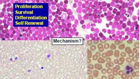
Cancer is the deadliest disease of children and leukemia is the most common childhood cancer. We are interested in the fundamental question how normal blood development is diverted into leukemia. What are the genetic and biochemical abnormalities that block cell differentiation, enhance proliferation and survival and confer the unique stem cell properties of self renewal to leukemia stem cells? We focus on chromosome 21 because of the mysterious association of leukemia with Down Syndrome. We utilize advanced genomic technologies, cell based assays of transformation of primary human and mouse stem cells, mouse models including transgenic, transplantation and explants of human leukemia. Our recent discoveries of the major involvement of the TSLP-IL7R-JAK2 pathway in leukemogenesis have lead to clinical trials with novel inhibitors of this pathway for high-risk leukemias in children and adults. The spread of leukemia to the brain is a major clinical problem as preventive therapy to the brain consistingof chemotherapy or irradiation causes long term side effects. We are therefore studying how leukemia cells spread to the central nervous system and developing mouse models to study this challenging problem.


We have discovered that SIL, a gene cloned from childhood leukemia, is required for centrosomal biogenesis and for survival of cancer cells. Targeting SIL by siRNA cause cancer cell death at mitotic entry in-vitro and in-vivo. Current research focuses on the fundamental role of the SIL protein in centrosome generation in normal and malignant cells and on developing approaches for its targeting for cancer therapy. 


Proteomics of Breast Cancer Progression

Research

Jun 22nd, 2015
Proteomics of Breast Cancer Progression

Prof. Tamar Geiger, Ph.D.
Department of Human Molecular Genetics and Biochemistry
Our main interest is to understand the mechanisms of breast cancer progression. We are using state-of-theart mass spectrometry-based proteomics to obtain a system-wide view of the tumor proteins. Analysis of the changes in protein levels and modifications that occur during tumor development is aimed to discover novel regulators of transformation.


Combination of the proteomics technology with biochemical and genetic methods will show the significance of these candidates to cancer development and may suggest novel drug targets and tumor markers.


Tel Aviv University makes every effort to respect copyright. If you own copyright to the content contained
here and / or the use of such content is in your opinion infringing Contact us as soon as possible >>